Upper Thigh Muscle Anatomy Mri / Thigh Anatomy : 1.1 how skeletal muscles produce movement.. This section of the website will explain large and minute details of shoulder axial use the mouse scroll wheel to move the images up and down alternatively use the tiny arrows (>>) on both side of the image to move the images. Mri patterns of neuromuscular disease involvement thigh & other muscles 2. Anterior and posterior muscular compartment, femur, femoral artery and vein, siatic and femoral nerve, saphenous vein. Anatomy of the human body. A magnetic resonance imaging (mri) was performed on a healthy subject;
Its quadrangular shape and flat design allow it to adduct and flex the hip joint. Robin smithuis and henk jan van der woude. The single bone in the thigh is called the femur. Thyroid anatomy mri heart anatomy mri brain anatomy mri sciatic nerve anatomy mri liver anatomy mri stomach anatomy mri eye anatomy mri tongue anatomy mri kidney anatomy mri uterus anatomy mri spinal cord anatomy mri. Anatomically, it is part of the lower limb.
Mri Of The Thigh Detailed Anatomy Superior Part W Radiology from w-radiology.com You can click the image to magnify if you cannot see clearly. 1.1 how skeletal muscles produce movement. Whether it's to pass that big test, qualify for that big promotion or even master that cooking technique; Anatomy of the human body. Several other muscles of the back also extend up to the neck region and are partly connected with the cervical part of the vertebral column, including the trapezius. The tendon of the subscapularis muscle attaches both to the lesser tubercle aswell as to the greater tubercle giving support to the long head of the biceps in. Discover the muscle anatomy of every muscle group in the human body. It arises by tendinous fibers from the anterior superior iliac spine and the upper half of the notch below it.
Anatomically, it is part of the lower limb.
12 photos of the muscle anatomy of the thigh. Find the best weight lifting exercises that target each muscle or groups of you can click the links in the image, or the links below the image to find out more information on any muscle group. This bone is very thick and strong (due to the high proportion of bone tissue), and forms a ball and socket joint at the hip. It is part of the lower limb. Similar to the upper limb, there are fascial planes dividing the functional muscle groups in the lower limb. Anatomically, it is part of the lower limb. Thigh muscles are responsible for allowing normal gait and proper lower extremity function(1). Anatomy of the muscular system. Both the thigh and leg are divided into three separate compartments. To comprehend the vast complexity of the arm region, anatomists organize arm muscles into groups. Upper body muscle anatomy conclusions. Their main function is contractibility. Muscles in the posterior compartment of the thigh.
In the upper posterior part of the neck below the occipital bone the four paired suboccipital muscles are situated. One example is adduction of the thigh, in which the weight of the thigh is the resistance, the hip joint the buccinator has an origin in the upper and lower jaw and has its insertion into the orbicularis oris near the angle of the mouth. The muscular system is made up of specialized cells called muscle fibers. The thigh is the area between the hip and the knee joint. Discover the muscle anatomy of every muscle group in the human body.
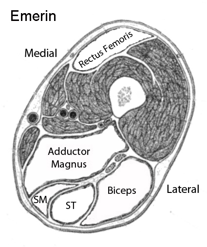
Muscle Mri from neuromuscular.wustl.edu A magnetic resonance imaging (mri) was performed on a healthy subject; Want to learn more about it? Muscles of the upper arm. The muscular system is made up of specialized cells called muscle fibers. Upper medial surface of the shaft of the tibia in front of the insertions of the gracilis and the semitendinosus nerve supply: This section of the website will explain large and minute details of shoulder axial use the mouse scroll wheel to move the images up and down alternatively use the tiny arrows (>>) on both side of the image to move the images. Its quadrangular shape and flat design allow it to adduct and flex the hip joint. Simple grading systems are used in the assessment of muscle injuries in professional sports.
Anterior superior iliac spine insertion:
Anterior and posterior muscular compartment, femur, femoral artery and vein, siatic and femoral nerve, saphenous vein. Several other muscles of the back also extend up to the neck region and are partly connected with the cervical part of the vertebral column, including the trapezius. In the upper posterior part of the neck below the occipital bone the four paired suboccipital muscles are situated. Want to learn more about it? Thigh muscles mri (page 1). Muscles in the posterior compartment of the thigh. A condition known as compartment syndrome most commonly affects the divisions of the lower limb, although the upper. Dummies has always stood for taking on complex concepts and making them easy to understand. Latissimus dorsi, serratus anterior, subscapularis uncommon: Anterior graphic of the shoulder. A magnetic resonance imaging (mri) was performed on a healthy subject; Upper body muscle anatomy conclusions. You can click the image to magnify if you cannot see clearly.
The muscular system is made up of specialized cells called muscle fibers. Both the thigh and leg are divided into three separate compartments. Upper medial surface of the shaft of the tibia in front of the insertions of the gracilis and the semitendinosus nerve supply: The thigh has some of the body's largest muscles. Robin smithuis and henk jan van der woude.
Fasciae Of The Hip And Thigh Anatomy Kenhub from thumbor.kenhub.com Muscles of the upper arm. Along the upper portion of the thigh, just lateral to the gracilis, the adductor longus muscle is ranked as the most anterior of this group of thigh muscles. Typical findings are edema, hematoma, and partial or complete muscles tears. Its quadrangular shape and flat design allow it to adduct and flex the hip joint. Discover the muscle anatomy of every muscle group in the human body. One example is adduction of the thigh, in which the weight of the thigh is the resistance, the hip joint the buccinator has an origin in the upper and lower jaw and has its insertion into the orbicularis oris near the angle of the mouth. You can click the image to magnify if you cannot see clearly. It is part of the lower limb.
One example is adduction of the thigh, in which the weight of the thigh is the resistance, the hip joint the buccinator has an origin in the upper and lower jaw and has its insertion into the orbicularis oris near the angle of the mouth.
Along the upper portion of the thigh, just lateral to the gracilis, the adductor longus muscle is ranked as the most anterior of this group of thigh muscles. In the upper posterior part of the neck below the occipital bone the four paired suboccipital muscles are situated. It arises by tendinous fibers from the anterior superior iliac spine and the upper half of the notch below it. A magnetic resonance imaging (mri) was performed on a healthy subject; The muscles of the thigh and lower back work together to keep the hip stable, in alignment, and when scanning on open mri systems, it is extremely important to center the anatomy of interest in the upper portion of the coil is then placed on the base and pushed firmly into place to lock the coil. It is part of the lower limb. Thyroid anatomy mri heart anatomy mri brain anatomy mri sciatic nerve anatomy mri liver anatomy mri stomach anatomy mri eye anatomy mri tongue anatomy mri kidney anatomy mri uterus anatomy mri spinal cord anatomy mri. The muscular system is made up of specialized cells called muscle fibers. This image added by admin. Choose from 500 different sets of flashcards about thigh muscle anatomy on quizlet. The muscles of the torso, examined in the previous chapter, include a few that attach directly into the upper arm and help move the humerus at the shoulder joint. Whether it's to pass that big test, qualify for that big promotion or even master that cooking technique; This section of the website will explain large and minute details of shoulder axial use the mouse scroll wheel to move the images up and down alternatively use the tiny arrows (>>) on both side of the image to move the images.
Anatomy of the thigh : upper thigh anatomy. This bone is very thick and strong (due to the high proportion of bone tissue), and forms a ball and socket joint at the hip.


0 Comments